Your nose is part of your respiratory system. It allows air to enter your body, then filters debris and warms and moistens the air. Your nose gives you a sense of smell and helps shape your appearance. Many common symptoms affect your nose, such as a stuffy nose and nosebleed. Other symptoms may need treatment to keep your nose functioning well. For more information visit Pritish Halder page.
Your nose, a structure that sticks out from the middle of your face, is part of your respiratory system as one of the sense organs.
The function of nose
Your nose is involved in several important bodily functions:
- Allows air to enter your body.
- Contributes to how you look and how you sound when you speak.
- Filters and cleans air to remove particles and allergens.
- Provides a sense of smell.
- Warms and moistens air so it can move comfortably into your respiratory system.
- Your nose is also a prominent aspect of your facial appearance and your sense of well-being.

Anatomy of human nose
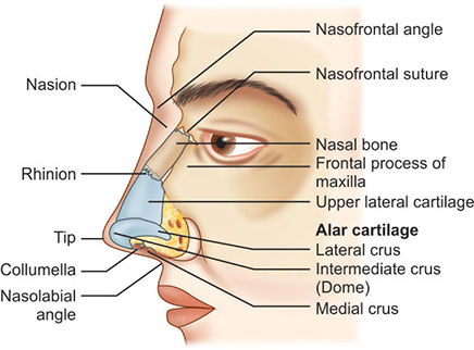
The external nose
The external nose is said to have a pyramidal shape. The nasal root is located superiorly, and is continuous with the forehead. The apex of the nose ends inferiorly in a rounded ‘tip’. Spanning between the root and apex is the dorsum of the nose.
Located immediately inferiorly to the apex are the nares; piriform openings into the vestibule of the nasal cavity. The nares are bounded medially by the nasal septum, and laterally by the ala nasi (the lateral cartilaginous wings of the nose).
Skeletal Structure
The skeleton of the external nose is made of both bony and cartilaginous components:
Bony component – located superiorly, and is comprised of contributions from the nasal bones, maxillae and frontal bone.
Cartilaginous component – located inferiorly, and is comprised of the two lateral cartilages, two alar cartilages and one septal cartilage. There are also some smaller alar cartilages present.
Whilst the skin over the bony part of the nose is thin, that overlying the cartilaginous part is thicker with many sebaceous glands. This skin extends into the vestibule of the nose via the nares. Here there are hairs which function to filter air as it enters the respiratory system.
Muscles
A number of small muscles insert into the external nose, contributing to facial expression. All these muscles are innervated by branches of the facial nerve .
The procerus muscle originates in the fascia overlying the nasal bone and lateral nasal cartilage, inserting into the inferior forehead. Contraction can depress the medial eyebrows, and wrinkles the skin of the superior dorsum.
The transverse portion of the nasalis muscle assists the procerus muscle in this action. Meanwhile the alar part of nasalis arises from the maxilla, inserting into the major alar cartilage. This allows the muscle to dilate the nares, “flaring” them. This action is assisted by the depressor septi nasi.
Vessels and Lymphatics
The skin of the external nose receives arterial supply from branches of the maxillary and ophthalmic arteries. The septum and alar cartilages receive additional supply from the angular artery and lateral nasal artery. These are both branches of the facial artery (derived from the external carotid artery).
Venous drainage is into the facial vein, and then in turn into the internal jugular vein.
Lymphatic drainage from the external nose is via superficial lymphatic vessels accompanying the facial vein. These vessels, like all lymphatic vessels of the head and neck, ultimately drain into the deep cervical lymph nodes.
The nasal cavity
The nasal cavity is the most superior part of the respiratory tract. It extends from the vestibule of the nose to the nasopharynx, and has three divisions:
- Vestibule – the area surrounding the anterior external opening to the nasal cavity.
- Respiratory region – lined by a ciliated psudeostratified epithelium, interspersed with mucus-secreting goblet cells.
- Olfactory region – located at the apex of the nasal cavity. It is lined by olfactory cells with olfactory receptors.
Nasal Conchae
Projecting out of the lateral walls of the nasal cavity are curved shelves of bone. They are called conchae (or turbinates). The are three conchae – inferior, middle and superior.
- Inferior meatus – between the inferior concha and floor of the nasal cavity.
- Middle meatus – between the inferior and middle concha.
- Superior meatus – between the middle and superior concha.
- Spheno-ethmoidal recess – superiorly and posteriorly to the superior concha.
The function of the conchae is to increase the surface area of the nasal cavity – this increases the amount of inspired air that can come into contact with the cavity walls. They also disrupt the fast, laminar flow of the air, making it slow and turbulent. The air spends longer in the nasal cavity, so that it can be humidified.
Openings into the Nasal Cavity
One of the functions of the nose is to drain a variety of structures. Thus, there are many openings into the nasal cavity, by which drainage occurs.
The paranasal sinuses drain into the nasal cavity. The frontal, maxillary and anterior ethmoidal sinuses open into the middle meatus. The location of this opening is marked by the semilunar hiatus, a crescent-shaped groove on the lateral walls of the nasal cavity.
The middle ethmoidal sinuses empty out onto a structure called the ethmoidal bulla. This is a bulge in the lateral wall formed by the middle ethmoidal sinus itself. The posterior ethmoidal sinuses open out at the level of the superior meatus.
The only structure not to empty out onto the lateral walls of the nasal cavity is the sphenoid sinus. It drains onto the posterior roof.In addition to the paranasal sinuses, other structures open into the nasal cavity:
Nasolacrimal duct – acts to drain tears from the eye. It opens into the inferior meatus.
Auditory (Eustachian) tube – opens into the nasopharynx at the level of the inferior meatus. It allows the middle ear to equalise with the atmospheric air pressure.
Gateways to the Nasal Cavity
As well as openings for the drainage of structures, nerves, vasculature and lymphatics need to be able to access the nasal cavity.
The cribriform plate is part of the ethmoid bone. It forms a portion of the roof of the nasal cavity. It contains very small perforations, allowing fibres of the olfactory nerve to enter and exit,
At the level of the superior meatus, the sphenopalatine foramen is located. This hole allows communication between the nasal cavity and the pterygopalatine fossa. The sphenopalatine artery, nasopalatine and superior nasal nerves pass through here.
The incisive canal is a pathway between the nasal cavity and the incisive fossa of the oral cavity. It transmits the nasopalatine nerve and greater palatine artery.
Vasculature
The nose has a very rich vascular supply – this allows it to effectively change humidity and temperature of inspired air. The nose receives blood from both the internal and external carotid arteries:
Internal carotid branches:
- Anterior ethmoidal artery
- Posterior ethmoidal artery
The ethmoidal arteries are branch of the ophthalmic artery. They descend into the nasal cavity through the cribriform plate.
External carotid branches:
- Sphenopalatine artery
- Greater palatine artery
- Superior labial artery
- Lateral nasal arteries
In addition to the rich blood supply, these arteries form anastomoses with each other. This is particularly prevalent in the anterior portion of the nose .The veins of the nose tend to follow the arteries. They drain into the pterygoid plexus, facial vein or cavernous sinus.
In some individuals, a few nasal veins join with the sagittal sinus (a dural venous sinus). This represents a potential pathway by which infection can spread from the nose into the cranial cavity.


